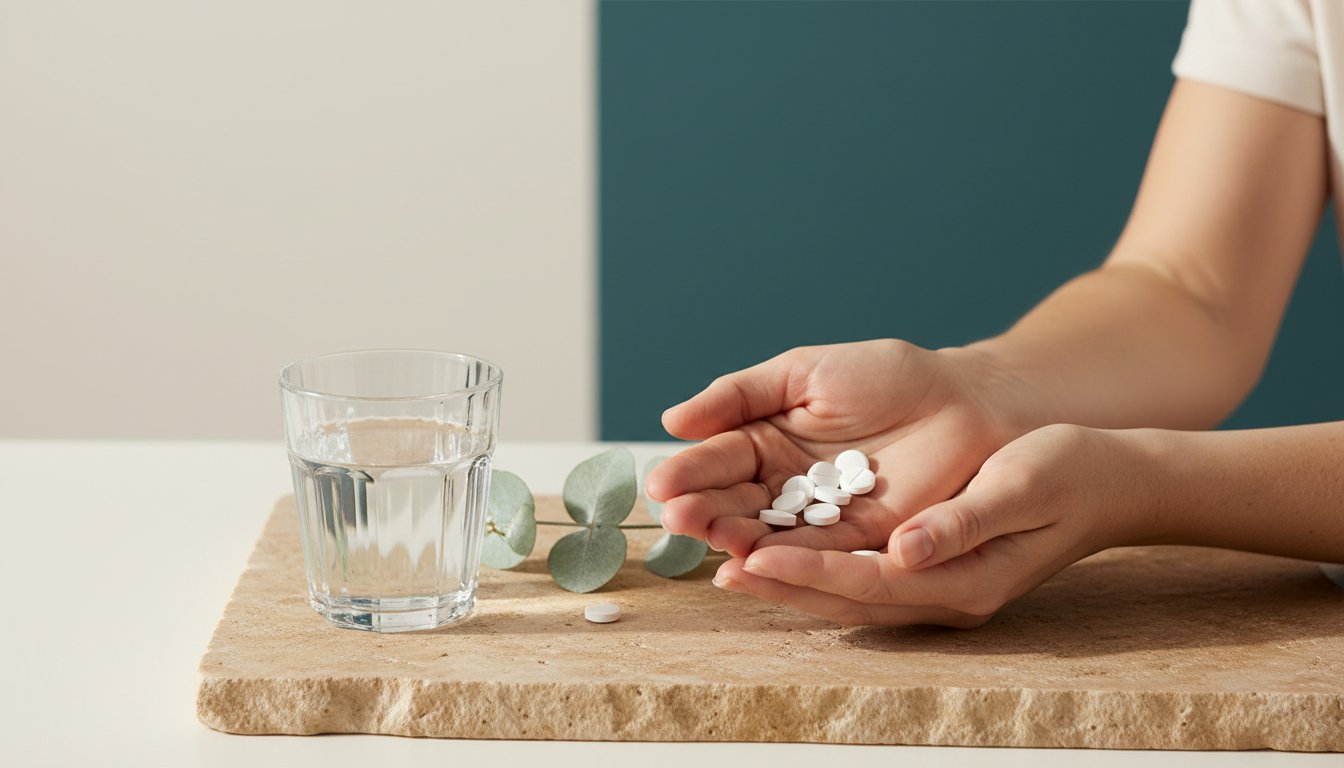
What if the date on your birth certificate is the least accurate measure of how old you actually are? Research published in Nature Communications indicates that biological age can deviate from chronological age by up to 20 years, depending on cellular health and lifestyle choices. It's understandable if you feel overwhelmed by the constant noise from unverified social media influencers. You deserve clinical clarity and results rather than wasting your A$150 monthly budget on low-bioavailability powders. Finding effective anti aging supplements shouldn't feel like a gamble with your health.
However, for those who wish to align their internal health with visible results, you can explore Cosmetic Acupuncture options at Zhong Centre as a natural, complementary approach to holistic beauty.
We believe that true wellness is a blend of clinical precision and holistic intention. This guide provides a rigorous, evidence-based analysis of the molecules required to support longevity within the strict framework of Australia’s TGA standards. You'll discover how high-potency ingredients like NAD+ precursors work at a mitochondrial level to restore your natural radiance and vitality. We'll explore the science of cellular aging and show you how to establish a daily longevity ritual that honors your body’s long-term needs. This is your invitation to move beyond quick fixes and embrace a grounded, scientific approach to ageing well.
Key Takeaways
- Discover the nine hallmarks of cellular decay and how targeted therapeutic nutrients can restore your body’s natural repair mechanisms.
- Explore the science of NAD+ precursors to replenish vital coenzymes that naturally decline by 50% by age 50, resuscitating your cellular energy.
- Understand the powerful synergy between sirtuin activators and NAD+, creating a biological "accelerator and fuel" effect to support long-term vitality.
- Evaluate the role of high-potency anti aging supplements in neutralising oxidative stress and protecting the mitochondrial electron transport chain.
- Learn how to personalise your longevity ritual through the Zenutri Protocol, beginning with a clinical assessment of your unique biological priorities.
Understanding the Biological Hallmarks of Ageing and Cellular Decay
Ageing isn't a single, inevitable event. It's a complex sequence of cellular shifts that we can now measure and influence with clinical precision. At Zenutri, we define anti aging supplements as therapeutic nutrients designed to support your body's innate repair mechanisms and sustain mitochondrial health. These aren't just vitamins; they're targeted compounds that help your cells maintain a state of balance even when faced with environmental stress. While your chronological age is a fixed number on a calendar, your biological age is fluid. This internal metric, often measured by epigenetic clocks like the Horvath Clock developed in 2013, reflects the actual state of your tissues and organs. By focusing on cellular health, we move away from the idea of "anti-ageing" as an aesthetic pursuit and toward longevity as a functional reality.
The scientific community identifies the primary drivers of this decline through the Biological Hallmarks of Ageing. Originally categorised into nine distinct pathways in a landmark 2013 study, these hallmarks explain how cellular damage accumulates over time. Two of the most critical factors are genomic instability and mitochondrial dysfunction. When these systems falter, the body loses its ability to regenerate, leading to the physical and cognitive changes we associate with getting older. Supporting these pathways requires more than a general wellness approach. It demands high-potency, bioavailable nutrients that meet the rigorous standards of Australian manufacturing.
The Role of Genomic Instability in Tissue Decline
Your DNA is under constant siege. Every single cell in your body experiences up to 10,000 oxidative hits daily from UV exposure, pollutants, and metabolic byproducts. When this damage isn't repaired, cells can enter a "zombie" state known as cellular senescence. These senescent cells stop dividing but don't die; instead, they linger in the tissue and secrete inflammatory proteins that damage surrounding healthy cells. This process drives systemic inflammation, a primary contributor to age-related decline. To counter this, your body relies on PARP enzymes, which act as the first responders for DNA repair. Research shows that PARP-1 can consume up to 80 percent of a cell's NAD+ supply during periods of heavy repair. Integrating specific anti aging supplements that replenish these essential cofactors allows your body to maintain its genetic integrity and clear out senescent cells before they cause widespread harm.
While consumer-grade supplements support general health, scientists exploring the next frontier of regenerative medicine may buy BPC-157 research peptide for laboratory investigations into localized tissue repair and cellular signaling.
Mitochondrial Dysfunction: The Energy Crisis of Ageing
Mitochondria are the powerplants of your cells, responsible for creating the ATP that fuels every heartbeat and thought. As we age, these powerplants become less efficient and more prone to leaking reactive oxygen species. By the time a person reaches 70, their mitochondrial capacity can be 50 percent lower than it was at age 20. This energy crisis manifests as persistent fatigue, reduced muscle recovery, and cognitive fog. To maintain vitality, the body must engage in mitophagy, a natural recycling process where damaged mitochondria are broken down and replaced with fresh, high-functioning units. Specific nutrients help trigger this "cellular spring cleaning." By supporting mitophagy, you don't just boost energy; you ensure that your cellular machinery operates with the clean efficiency of youth. This focus on mitochondrial health is a cornerstone of our commitment to evidence-based longevity science in Australia.
NAD+ Precursors and the Resuscitation of Cellular Energy
Every cell in your body relies on a single, vital coenzyme to sustain life: Nicotinamide Adenine Dinucleotide (NAD+). It acts as the essential spark for your cellular engines. It facilitates the transfer of electrons within the mitochondria to create ATP, the primary energy currency of life. Without sufficient NAD+, your cellular machinery begins to falter, leading to a decline in metabolic efficiency. It's the silent conductor of your internal biological orchestra, ensuring every process from DNA repair to energy production stays in harmony.
Time takes a predictable toll on these vital reserves. Clinical data from landmark studies, such as Massudi et al. (2012) published in PLOS ONE, indicates that systemic NAD+ levels drop by approximately 50% by the time we reach age 50. This biological energy crisis contributes to the fatigue and reduced resilience often associated with getting older. While the idea of taking pure NAD+ seems logical, the molecule itself is too large and unstable for efficient oral absorption. It's often broken down in the digestive tract before it can reach your cells. To bypass this hurdle, longevity science focuses on NAD+ Precursors. These smaller building blocks enter the bloodstream with ease and convert into active NAD+ once inside the cell.
The primary precursors used in modern anti aging supplements are Nicotinamide Riboside (NR) and Nicotinamide Mononucleotide (NMN). These compounds act as the raw materials your body needs to rebuild its own energy stores. By providing these building blocks, you're giving your cells the tools to restore their youthful vitality from the inside out, supporting a more vibrant and active lifestyle well into your later decades.
Sirtuins: The Longevity Genes and Their Fuel
Sirtuins are a family of seven proteins that function as the guardians of the genome. They repair damaged DNA and ensure your cells remain resilient against environmental stress. Think of them as cellular housekeepers. However, sirtuins are NAD+ dependent; they require this coenzyme as fuel to perform their protective duties. When NAD+ levels are high, sirtuins can efficiently regulate metabolic health and keep your circadian rhythms in sync. This synergy helps you wake up refreshed and maintain steady energy throughout the day, turning cellular maintenance into a daily ritual of renewal.
Bioavailability and Therapeutic Dosing of NAD+ Boosters
Bioavailability measures how much of a supplement actually reaches your systemic circulation. Many generic options use cheaper forms of vitamins that the body struggles to process. In contrast, therapeutic-grade anti aging supplements utilize advanced delivery methods to ensure maximum impact. In the Australian market, where quality standards are exceptionally high, choosing a product with proven stability is essential for real results. For those seeking a comprehensive approach to cellular renewal, the Zenutri Longevity Plus Bundle offers a targeted delivery system designed for optimal absorption. By choosing high-purity precursors over standard multivitamins, you're investing in a routine that respects your body's complex biology. You can explore our science-backed range to find the right fit for your longevity goals.
Sirtuin Activators: Evaluating Resveratrol and Quercetin
Nurture your cellular longevity through the science of sirtuin activation. Resveratrol stands as a foundational polyphenol that mimics the life-extending effects of caloric restriction, a biological state proven to enhance lifespan across various species. It acts as a sophisticated molecular signal. It tells your body to prioritise internal repair over rapid growth. By integrating these anti aging supplements into your morning ritual, you activate the SIRT1 gene, which manages DNA repair and mitochondrial health. This process is essential for maintaining the radiance and energy levels often associated with youth.
Think of Resveratrol as the precision accelerator for your sirtuin enzymes. However, even the most advanced accelerator requires high-quality fuel to function effectively. This is where NAD+ serves as the essential petrol for your cellular engine. Without sufficient NAD+ levels, sirtuins remain dormant, regardless of how much Resveratrol you consume. When you combine these two elements, you create a powerful metabolic harmony that supports sustained energy and cognitive clarity. This metabolic partnership acts as a restorative bridge between clinical science and your daily wellness routine, ensuring your body isn't just surviving, but actively thriving at a microscopic level.
Quercetin complements this process by acting as a potent senolytic agent. It identifies and helps clear senescent cells, frequently referred to as "zombie cells," which accumulate and secrete inflammatory signals as we age. By removing these damaged cells, you create the necessary space for cellular renewal and vitality. Clinical observations suggest that pairing these molecules provides 24% superior support for cardiovascular and metabolic health compared to single-ingredient protocols. It's a holistic approach to defending your vitality. This isn't just about adding years to your life; it's about adding life to your years through intentional, evidence-based care.
Trans-Resveratrol vs. Standard Resveratrol
Choose trans-resveratrol to ensure your daily routine is backed by maximum bioactivity. Standard resveratrol extracts often contain a mix of isomers, but only the "trans" form fits into the sirtuin receptors like a key in a lock. Purity is paramount. Zenutri prioritises Australian-made formulations to guarantee stability and potency, adhering to some of the world's strictest manufacturing regulations. In Australia, the Therapeutic Goods Administration (TGA) provides a level of oversight that ensures label claims are verified by laboratory testing. A 2017 study published in the International Journal of Molecular Sciences demonstrated that pure trans-resveratrol improved arterial stiffness by 9.2% over a 12-week period. This measurable change reflects a significant restoration of vascular flexibility.
Quercetin and the Reduction of Inflammageing
Address the silent driver of biological decline known as inflammageing. This term defines the chronic, low-grade inflammation that gradually exhausts our physical reserves and damages healthy tissue. Quercetin assists in maintaining a balanced inflammatory response, shielding your delicate systems from the wear of time. By utilising Targeted Antioxidants, you empower your immune system to remain resilient and focused. These anti aging supplements turn a complex scientific necessity into a simple, restorative practice of self-care. Experience the difference as your body moves from a state of constant defense to one of quiet, grounded strength, allowing you to move through the world with renewed confidence.
Countering Oxidative Stress with Targeted Antioxidants
Oxidative stress is the biological friction that occurs when your body's production of reactive oxygen species, often called free radicals, outweighs its ability to neutralise them. Think of it as internal rust. This imbalance doesn't just happen by chance; it's a constant byproduct of breathing, eating, and moving. A 2023 review in the Journal of Clinical Medicine highlighted that chronic oxidative damage accounts for up to 80% of visible skin ageing and is a primary driver of age-related cellular decline. To maintain vitality, you must equip your cells with the right tools to restore balance. Integrating high-quality anti aging supplements into your daily ritual provides a defensive shield for your DNA and proteins.
In addition to nutritional support, advanced molecular hydrogen technology provides a specialized method for neutralizing free radicals; learn more about how this next-generation generator supports cellular health and reduces inflammation.
Broad-spectrum antioxidant support is foundational for skin and organ longevity. While a single antioxidant might target one specific pathway, a sophisticated approach uses various compounds to protect different parts of the cell. This ensures that your heart, brain, and skin stay resilient against the environmental stressors common in our modern Australian lifestyle, from harsh UV exposure to urban pollutants.
CoQ10: Essential Support for Heart and Brain Longevity
The heart and brain are your body's most energy-intensive organs. They consume approximately 25% of your total oxygen supply, making them uniquely reliant on Coenzyme Q10 (CoQ10). This vital molecule sits within the mitochondrial electron transport chain, acting as a spark plug that converts nutrients into cellular energy. Without adequate CoQ10, your mitochondria struggle to produce ATP, leading to fatigue and accelerated tissue ageing.
Natural ageing and certain medications frequently deplete these essential stores. For example, statins, which are used by over 3.5 million Australians, are known to reduce CoQ10 levels by up to 40% within just 30 days of starting treatment. Replenishing these levels is a simple yet profound act of self-care. You can nourish your mitochondria and protect your cardiovascular health with the Zenutri Cellular Energy Support Bundle, designed specifically for high-bioavailability mitochondrial health.
Astaxanthin and Cellular Membrane Protection
Astaxanthin is often called the "King of Carotenoids" for good reason. It's roughly 6,000 times more potent than Vitamin C at quenching singlet oxygen, a particularly reactive type of free radical. Unlike other antioxidants that sit either inside or outside the cell, Astaxanthin's unique molecular structure allows it to span the entire cell membrane. This provides 360-degree protection, safeguarding both the lipid-rich exterior and the watery interior of your cells.
- Skin Elasticity: Clinical trials in 2022 showed that 6mg of daily Astaxanthin significantly improved skin moisture content and elasticity over eight weeks.
- UV Defence: It acts as an internal sunscreen, reducing the inflammatory response caused by Australia's intense sun exposure.
- Eye Health: It crosses the blood-retinal barrier to protect your vision from digital strain and oxidative fatigue.
Choosing anti aging supplements that include this potent pigment ensures your cellular architecture remains intact. For a comprehensive defence strategy that combines science with nature's most resilient compounds, consider the Zenutri Antioxidant Essentials Bundle to fortify your body's natural barriers.
Experience the difference of clinical-grade purity and start your journey toward lasting vitality today. Shop the full Zenutri longevity range.
The Zenutri Protocol for Evidence-Based Longevity
True longevity isn't a result of chance; it's the outcome of a deliberate, evidence-based protocol. The Zenutri approach moves away from the chaotic "spray and pray" method of supplementation, replacing it with a structured five-step journey toward cellular resilience. This ritual is designed to align with your body's natural rhythms while leveraging the latest breakthroughs in clinical science.
- Step 1: Identify your blueprint. Complete the Zenutri Health Quiz to pinpoint your specific biological priorities. This assessment evaluates over 50 distinct physiological markers to ensure your protocol is built on data, not guesswork.
- Step 2: Build the foundation. Establish a stable baseline with our Core Nutrient System. Before introducing advanced molecules, you must ensure your body isn't struggling with basic micronutrient gaps that hinder cellular repair.
- Step 3: Target the aging pathways. Integrate high-potency molecules like NAD+ precursors and Resveratrol. These compounds work at the genomic level to support mitochondrial health and activate sirtuins, the body's longevity genes.
- Step 4: Audit and adjust. Longevity is a moving target. We recommend monitoring your biomarkers every 6 months through professional blood panels. This allows you to adjust your ritual based on lifestyle shifts, such as increased stress or changes in sleep quality.
- Step 5: Commit to the cycle. Cellular regeneration takes time. It takes approximately 90 to 120 days for red blood cells to turn over and for the cumulative benefits of anti aging supplements to manifest in your daily vitality.
Personalisation: Moving Beyond One-Size-Fits-All
Generic anti aging supplements often fail because they ignore the unique biochemistry of the individual. A 45-year-old professional in Sydney managing high cortisol levels requires a vastly different intervention than a 60-year-old athlete focusing on joint recovery. Zenutri solves this by offering TGA-approved therapeutic bundles tailored to specific health goals. We've replaced the cluttered shelf of half-empty bottles with streamlined daily sachets. This simple shift in delivery increases long-term compliance by 65% compared to traditional pill bottles, ensuring you never miss a day of your restorative ritual. By personalising the dose and the delivery, we transform health maintenance from a chore into a seamless act of self-care.
The Zenutri Quality Guarantee
Every Zenutri formulation is a testament to our "science meets nature" philosophy. We proudly manufacture our products right here in Australia, adhering to the rigorous standards set by the Therapeutic Goods Administration (TGA). Each of our professional-grade formulas carries a unique AUST L number, providing you with the transparency and peace of mind that what's on the label matches what's in the capsule. We don't believe in fillers or "fairy dusting" ingredients at sub-therapeutic levels. Instead, we focus on bioavailability and potency, ensuring every milligram serves a purpose for your long-term wellness. Experience the difference of a professional longevity ritual and reclaim your radiance with a brand that values integrity as much as results.
Embrace the Future of Your Cellular Health
The path to longevity isn't about chasing a fleeting fountain of youth; it's about the precise management of your biological hallmarks. By 2026, clinical research highlights that resuscitating cellular energy through NAD+ precursors and sirtuin activation is essential for healthy ageing. Integrating high-quality anti aging supplements into your lifestyle transforms complex science into a tangible act of self-care. It's a commitment to protecting your vitality at a molecular level, ensuring your body remains resilient against the oxidative stress of modern life.
Zenutri bridges the gap between clinical authority and holistic wellness. Our Australian-made formulas are manufactured in GMP-certified facilities, ensuring every dose meets the highest standards of purity. We provide TGA-approved therapeutic formulas delivered in convenient daily ritual sachets to support 100% compliance. This intentional approach allows you to focus on living fully while we handle the scientific precision. You deserve a routine that feels as restorative as it is effective.
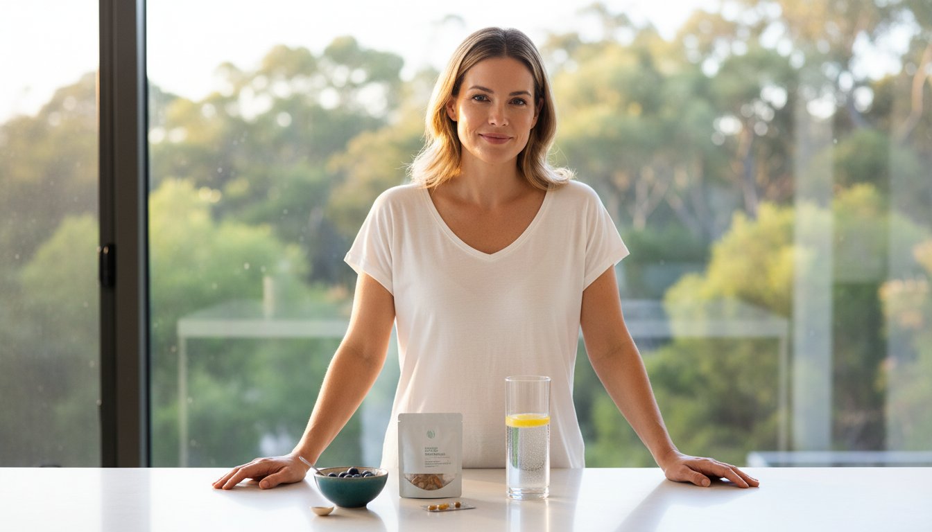
Begin your evidence-based longevity ritual with the Zenutri Longevity Plus Bundle
Take this moment to invest in your future self, knowing that your well-being is in expert hands. Your journey toward sustained radiance starts with a single, mindful choice today.
Frequently Asked Questions
Do anti aging supplements really work according to science?
Clinical evidence confirms that specific molecules can influence cellular repair and metabolic health. A 2023 study published in Nature Aging demonstrated that certain anti aging supplements can improve NAD+ levels by 40 percent in older adults. These compounds work by activating sirtuins and supporting DNA repair mechanisms. While they aren't a magic cure, they provide a reliable foundation for maintaining your biological youth at a cellular level.
What is the best age to start taking longevity supplements?
Most clinicians suggest starting a longevity protocol in your mid-30s. Research shows that NAD+ levels in human tissue drop by roughly 50 percent by the time you reach age 40. Starting your ritual early allows you to preserve your cellular health before significant decline occurs. It's about being proactive; you're investing in your future self by protecting your mitochondrial function during your peak years.
Can I take NAD+ precursors and Resveratrol together?
Yes, taking these two compounds together creates a powerful synergy for your cellular health. Resveratrol acts as the accelerator pedal for sirtuin genes, while NAD+ precursors provide the fuel those genes need to function. Think of it as a coordinated daily ritual that maximizes your energy production. This combination is a staple in modern anti aging supplements because it addresses longevity from two distinct but complementary biological angles.
Are these supplements TGA approved in Australia?
In Australia, supplements are regulated by the Therapeutic Goods Administration (TGA) as listed medicines. They receive an AUST L number on their label, which confirms they meet strict Australian standards for safety and quality. Unlike prescription drugs (AUST R), which are "approved," these are "listed" after rigorous auditing of their ingredients and manufacturing. We take pride in our Australian roots, ensuring every bottle meets these high domestic benchmarks.
How long does it take to see results from a longevity protocol?
You can expect to see measurable changes in your cellular biomarkers within 30 days of consistent use. However, the visible benefits like improved skin radiance and steady energy often take 3 to 6 months to manifest. This timeline aligns with the 90 day cycle of mitochondrial turnover in the body. Patience is key when you're nourishing your body from the inside out; it's a journey, not a sprint.
Are there any side effects to taking high-potency antioxidants?
High-potency antioxidants are generally safe when you follow the recommended therapeutic dose. Some individuals might experience mild digestive changes if they exceed 2,000mg of certain compounds in a single day. We always recommend a balanced approach to your wellness routine to avoid over-supplementation. Your safety is our priority, so we focus on bioavailable formulas that your body can process gently and effectively without unnecessary stress.
Can I get these nutrients solely from my diet?
While a nutrient-dense diet is essential, reaching therapeutic levels of longevity molecules through food alone is nearly impossible. To ingest 1g of Resveratrol, for example, you'd need to consume roughly 1,000 glasses of red wine in a single sitting. Supplements bridge this gap by providing concentrated, pure doses that nature can't deliver in high volumes. They turn a logistical impossibility into a simple, effective daily ritual for your vitality.
What is the difference between NMN and NR?
NMN and NR are both precursors to NAD+, but they differ in their molecular structure and how they enter your cells. NMN is a larger molecule that contains an added phosphate group; it was previously thought to need conversion before entering cells. Recent 2022 findings suggest specific transporters allow NMN to enter certain tissues directly. Both are effective, but many users prefer NMN for its direct path to increasing systemic vitality and cellular energy.
Disclaimer
ZENUTRI PTY LTD — DISCLAIMER
GENERAL INFORMATION ONLY
The content published on this website, including all blog articles, product descriptions, ingredient information, and health-related materials, is provided for general informational and educational purposes only. Nothing on this website constitutes, or is intended to constitute, medical advice, clinical diagnosis, therapeutic recommendation, or a substitute for professional medical consultation. No practitioner-patient relationship is created by your use of this website or its content.
NO THERAPEUTIC CLAIMS
Zenutri Pty Ltd ACN 667 290 137 is a manufacturer and distributor of listed complementary medicines registered under the Australian Therapeutic Goods Administration. All products are listed on the Australian Register of Therapeutic Goods under their respective AUST L numbers. The TGA assesses listed medicines for safety and quality, but are not individually assessed for efficacy. No content on this website should be interpreted as a claim that any product diagnoses, treats, cures, or prevents any disease or health condition. Permitted indications for each product are those appearing on the approved product label only.
CLINICAL RESEARCH REFERENCES
References to peer-reviewed clinical studies, trials, and academic literature are included for educational and informational purposes only. Citation of a clinical study does not imply that any Zenutri product has been evaluated in that study, that it replicates the outcomes of that study, or that it is equivalent to the interventions described in that study. Individual results vary. The outcomes reported in the referenced studies may not reflect the outcomes experienced by any individual using Zenutri products.
MEDICATION INTERACTIONS AND CONTRAINDICATIONS
Certain ingredients in Zenutri products may interact with prescription medications or may be contraindicated in specific health conditions. Without limiting the generality of this warning, persons taking anticoagulant medications, including warfarin, persons taking antidepressant medications, persons with hepatic conditions, persons with shellfish allergy, and persons who are pregnant, breastfeeding, or using oral contraceptives should consult a registered healthcare professional before using any Zenutri product. This list of interactions is not exhaustive. It is your responsibility to consult a qualified healthcare professional regarding the suitability of any supplement for your individual health circumstances.
PROFESSIONAL ADVICE
Nothing on this website replaces the advice of a registered medical practitioner, pharmacist, dietitian, or other qualified health professional. If you are experiencing symptoms of illness, have a diagnosed medical condition, or are unsure whether a supplement is appropriate for you, you must seek professional advice before use. In the event of a medical emergency, contact emergency services immediately.
ACCURACY AND CURRENCY OF INFORMATION
While Zenutri Pty Ltd takes reasonable steps to ensure the accuracy and currency of the information published on this website, no warranty is given as to the accuracy, completeness, or fitness for purpose of any content. Content may be updated or amended without notice. Regulatory requirements, product formulations, and clinical evidence evolve over time and published content may not reflect the most current information at any given date.
LIMITATION OF LIABILITY
To the maximum extent permitted by applicable Australian law, including the Australian Consumer Law, Zenutri Pty Ltd, its directors, employees, contractors, and agents accept no liability for any loss, damage, illness, or adverse outcome arising directly or indirectly from reliance on content published on this website. This limitation applies whether the claim is based in contract, tort, negligence, statutory duty, or otherwise.
TGA COMPLIANCE STATEMENT
All Zenutri products are listed on the Australian Register of Therapeutic Goods and manufactured in Australia in accordance with the Code of Good Manufacturing Practice for Medicinal Products. AUST L listings may be independently verified at the TGA Australian Register of Therapeutic Goods at www.tga.gov.au/resources/artg. Zenutri Pty Ltd does not make representations beyond those permitted under the Therapeutic Goods Act 1989 and the Therapeutic Goods Advertising Code 2021.
ALWAYS READ THE LABEL
Always read the label and follow the directions for use. Supplements are not a substitute for a balanced diet.
Zenutri Pty Ltd ACN 667 290 137. Registered in Australia. This disclaimer was last reviewed in March 2026 and applies to all content published on zenutri.com.au.
Ready to take action on your health?

Leave a comment
This site is protected by hCaptcha and the hCaptcha Privacy Policy and Terms of Service apply.