
In 1992, Hungarian physician András Czeizel and his colleague Istvan Dudás published a randomised controlled trial in the New England Journal of Medicine that would become one of the most consequential single studies in the history of prenatal medicine. The trial enrolled 4,753 Hungarian women planning their first pregnancies and randomly assigned them to one of two groups: a multivitamin supplement containing 0.8mg folic acid taken from at least one month before conception through the second missed menstrual period, or a trace-element supplement containing no folate. The folate group had zero neural tube defects. The control group had six. The difference was statistically significant and represented a risk reduction of more than 70% for the most preventable category of birth defect — one that, in Australia alone, affects approximately 300 pregnancies per year even in an era when folate-enriched foods and preconception supplement awareness have substantially reduced but not eliminated the deficit. The mechanism was not mysterious: neural tube closure — the folding and fusion of the neural plate into the closed tube that becomes the brain and spinal cord — occurs between days 21 and 28 of gestation and requires a rate of cell division so rapid that thymidylate synthase (the enzyme producing the DNA building block dTMP from dUMP, using folate as its essential methyl donor) becomes the rate-limiting step in whether the closure proceeds without interruption. Inadequate folate means inadequate dTMP, inadequate cell division speed at the closure front, and a window of vulnerability that environmental and genetic factors can exploit. This one mechanism — thymidylate synthesis at the neural plate — is the most robustly evidence-based single rationale for any prenatal supplement recommendation in obstetric medicine.
The Czeizel trial is where the evidence for maternity supplements in Australia begins — but it is far from where it ends. The five components of the Zenutri Maternity Foundation Bundle each have mechanistic evidence bases of comparable depth: DHA's role in the third-trimester brain growth spurt and the specific phospholipid architecture of fetal neural membranes; iron's provision of both the expanded maternal haemoglobin mass and the fetal iron stores that must sustain an infant through months of iron-poor breast milk; iodine's continuation through the fourth trimester of breastfeeding during which the sodium/iodide symporter in mammary gland tissue actively concentrates iodine into milk for the neonate's continuing thyroid-dependent neurological development; and the D3-osteocalcin-K2 MK-7 axis that directs the third trimester's massive calcium accretion into the fetal skeleton rather than soft tissue while simultaneously preserving maternal bone density. This article provides the molecular evidence for all five — not as generic "prenatal nutrition is important" reassurance, but as the specific biological mechanisms that each formulation component directly addresses. Always confirm your full prenatal protocol with your GP, obstetrician, or midwife before initiating supplementation — these mechanisms inform the conversation; your healthcare provider determines the protocol.
Key Takeaways
- Understand the Czeizel 1992 NEJM folate RCT — the randomised controlled trial establishing greater than 70% neural tube defect risk reduction with periconceptional folate supplementation — and the thymidylate synthase mechanism that explains why folate's rate-limiting role in dTMP synthesis for DNA replication makes the 21-to-28-day neural tube closure window acutely sensitive to folate status, and why the preconception initiation timing that most Australian women miss is clinically essential rather than merely precautionary.
- Discover the third-trimester DHA brain growth spurt — the accelerated DHA accretion rate of approximately 30-40mg per day in fetal brain tissue during the third trimester, the specific phospholipid classes (phosphatidylserine and phosphatidylethanolamine in neuronal inner leaflet membranes, phosphatidylcholine in myelin) that concentrate DHA to determine membrane fluidity and synaptic transmission efficiency, and why the retinal phospholipid matrix's 50% DHA content in photoreceptor disc membranes makes visual development a co-dependent outcome of maternal DHA adequacy alongside neurological development.
- Learn the fetal haemoglobin oxygen delivery biology — how fetal haemoglobin (HbF, alpha₂gamma₂ tetramers) achieves higher oxygen affinity than adult HbA (alpha₂beta₂) through reduced 2,3-bisphosphoglycerate (2,3-BPG) binding to the gamma chain, creating the oxygen affinity differential that drives oxygen transfer from maternal to fetal circulation at the placenta — and why the iron required for HbF synthesis must be provided both during gestation and as a postpartum iron store sufficient to sustain the infant through the first 4-6 months when breast milk iron alone provides less than 15% of the infant's daily iron need.
- Understand fourth-trimester breastfeeding iodine biology — how the sodium/iodide symporter (NIS) in mammary gland epithelial cells actively concentrates iodine from maternal plasma into breast milk at a 20-50 fold concentration gradient, why breast milk iodine concentration is directly determined by maternal iodine status, and why the postpartum iodine requirement increases to 270mcg/day (above the 220mcg/day gestational level) making continued iodine supplementation through the breastfeeding period as clinically consequential as its preconception initiation.
- Apply the D3-osteocalcin-K2 MK-7 fetal skeletal calcium axis — how Vitamin D3 drives intestinal calcium absorption and circulating calcium bioavailability, how K2 MK-7 gamma-carboxylates osteocalcin (enabling hydroxyapatite binding in bone matrix) and matrix Gla protein (preventing soft-tissue and vascular calcification), why uncarboxylated osteocalcin from K2 insufficiency fails to direct the third trimester's approximately 30g fetal calcium accretion to the skeleton, and how the Knapen 2013 Osteoporosis International RCT validates the 180mcg MK-7 dose in Osteo+Core (AUST L 520792) — the TGA-listed anchor of the Zenutri Maternity Foundation Bundle at $55 AUD.
Folate, Neural Tube Closure, and the Czeizel 1992 Evidence
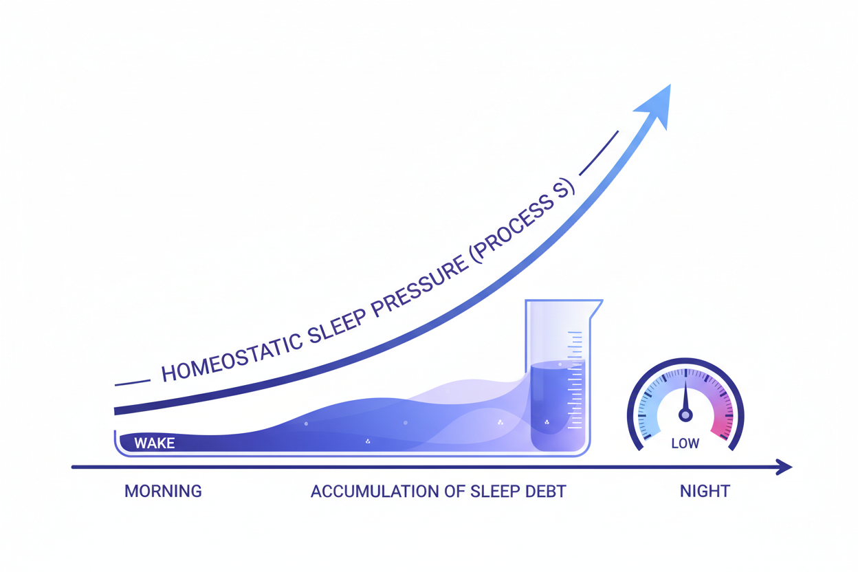
The thymidylate synthesis pathway is the molecular engine of cell division — the biochemical step that produces deoxyThymidine 5'-Monophosphate (dTMP), one of the four DNA building blocks, from deoxyUridine 5'-Monophosphate (dUMP) through the thymidylate synthase (TYMS) reaction that requires 5,10-methylenetetrahydrofolate as its methyl donor and that regenerates dihydrofolate as a by-product. Dihydrofolate must then be reduced back to tetrahydrofolate by dihydrofolate reductase (DHFR) to re-enter the folate cycle — a NADPH-consuming step that connects the entire pathway's flux to the cellular redox state. This is not a peripheral metabolic pathway. In a cell undergoing rapid division, dTMP production through TYMS is rate-limiting for the speed of DNA replication: inadequate 5,10-methyleneTHF substrate slows TYMS, reduces dTMP availability, creates uracil misincorporation into DNA (dUMP is incorporated in place of missing dTMP), and triggers DNA repair cycles that compete with the replication fork's forward progress. In the rapidly dividing neuroepithelial cells at the closing margins of the neural tube — cells that are dividing every 8-12 hours during the 21-to-28-day closure window — this folate dependency becomes the precise point of vulnerability that the Czeizel trial's zero-versus-six NTD result documents.
The clinical implication that the mechanism makes unavoidable is preconception initiation. Neural tube closure at day 21-28 occurs before most Australian women have taken a positive pregnancy test, visited a GP, or initiated prenatal supplementation. The RBC folate levels that reflect the tissue folate pool relevant to this closure window take 10-12 weeks of consistent supplementation to reach optimal concentrations — making a first-trimester prescription inadequate for the protection the Czeizel evidence demonstrates. Australian data consistently shows that fewer than 40% of women begin folate supplementation before conception, meaning the majority enter the most critical nutritional window for NTD prevention in a suboptimal folate state. The Zenutri Maternity Foundation Bundle's 500mcg Folate component addresses this with a dose consistent with Australian preconception guidance — above the minimum 400mcg and suitable for both the preconception and ongoing gestational periods. Take the free Zenutri health quiz to understand your nutritional baseline, and bring those results to your GP or obstetrician to confirm this and the remaining four components are appropriate for your individual health context.
MTHFR and the Active Form Question
The mechanism covered in the companion maternity supplements article explains why the form of folate in a prenatal supplement matters as much as the dose: the approximately 44% of Australians carrying C677T or A1298C MTHFR polymorphisms have 30-70% reduced MTHFR enzyme activity, impairing the conversion of 5,10-methyleneTHF to 5-MTHF (the methyl donor for homocysteine remethylation and DNA methylation) and allowing homocysteine accumulation that is independently associated with NTD risk beyond the direct thymidylate synthesis mechanism. The Zenutri Maternity Foundation Bundle includes Folate in its TGA-listed form — confirmed as the active folate compound for your specific formulation on the product label. Women with known MTHFR variants should discuss active folate form selection with their GP to confirm the product formulation meets their specific needs.
DHA, the Third-Trimester Brain Growth Spurt, and Fetal LCPUFA Accretion
The fetal brain does not grow at a uniform rate throughout gestation. It follows a staged developmental programme in which different processes — neuronal proliferation in the ventricular zone, neuronal migration to cortical layers, synaptogenesis, myelination, and glial cell expansion — occur in overlapping but temporally distinct waves, each with its own nutritional dependencies. The third trimester is characterised by a dramatic acceleration in all of these processes simultaneously — a "brain growth spurt" that takes the fetal brain from approximately 100g at 28 weeks to approximately 400g at term, representing a fourfold weight increase in the final 12 weeks of gestation driven primarily by the explosive expansion of synaptic connections, the proliferation of glial cells (particularly oligodendrocytes and astrocytes), and the onset of myelination in major white matter tracts. DHA — docosahexaenoic acid, a 22-carbon omega-6 with six double bonds — is the primary structural long-chain polyunsaturated fatty acid (LCPUFA) of the nervous system, and its accretion rate in fetal brain tissue peaks during precisely this third-trimester growth spurt.
Quantitative fatty acid analysis of postmortem human fetal and neonatal brain tissue — the experimental methodology that established the specific phospholipid composition of fetal neural membranes — shows DHA constituting approximately 14-17% of total fatty acids in the frontal cortex gray matter, and approximately 40% of the polyunsaturated fatty acid fraction. The structural role of DHA in neuronal membranes is not incidental: DHA's six double bonds create a highly flexible, low-melting-point acyl chain that maintains membrane fluidity at physiological temperatures, enabling the rapid conformational changes in membrane-embedded proteins — ion channels, receptor proteins, G-protein-coupled receptors, voltage-gated channels — that synaptic transmission requires. In the phosphatidylserine (PS) and phosphatidylethanolamine (PE) of neuronal inner-leaflet membranes, DHA is the preferred sn-2 position fatty acid. PS-DHA specifically recruits protein kinase C (PKC) to the membrane surface — a localization event required for the activation of the synaptic plasticity pathways that underlie memory formation. In retinal photoreceptor membranes, DHA constitutes approximately 50% of the fatty acids in rhodopsin-containing disc membranes, where its exceptional membrane fluidity enables the rapid conformational change of rhodopsin following photon absorption that initiates the phototransduction cascade. Australian dietary surveys consistently document DHA intake in pregnant women averaging 50-100mg/day — substantially below the 200mg/day required to support the third-trimester fetal accretion rate from maternal stores — making supplementation the necessary bridge between dietary reality and developmental requirement. The DHA 200-300mg component of the Zenutri Maternity Foundation Bundle addresses this directly, providing the dose range most consistently cited in current prenatal nutrition guidelines as adequate for fetal DHA status.
The Maternal DHA Store and Postpartum Depletion
The third-trimester fetal DHA accretion is not costless to the mother. Maternal plasma DHA concentrations decline progressively across pregnancy as the placenta preferentially transfers DHA to the fetal brain through a process of retroconversion and selective transport — a biologically programmed maternal sacrifice of DHA stores for fetal neurological development. Postpartum, breast milk DHA concentration is directly proportional to maternal DHA status (typically 0.2-0.3% of breast milk fatty acids in Australian women, lower than in populations with high fish consumption), providing the infant's primary DHA source during the first 6 months when retinal and hippocampal DHA accretion continue after birth. Continuing DHA supplementation through the breastfeeding period is supported by the same logic as the gestational period — and by the evidence that postpartum maternal DHA depletion contributes to the mood dysregulation and cognitive changes of the postpartum period, for which low omega-3 status is a proposed contributing mechanism. The Maternity Foundation Bundle's DHA component is appropriate for this continued postpartum use, with GP confirmation recommended.
Iron, Fetal Haemoglobin Oxygen Delivery, and the Postnatal Iron Store
The oxygen delivery from mother to fetus across the placenta depends on a precise and elegant biochemical mechanism: the oxygen affinity differential between maternal haemoglobin and fetal haemoglobin that drives oxygen transfer in the direction of the developing life even when maternal oxygen partial pressure is only moderately above fetal levels. Adult haemoglobin (HbA) is an alpha₂beta₂ tetramer — two alpha and two beta globin chains, each with an iron-containing haem prosthetic group that binds oxygen reversibly at its ferrous (Fe²⁺) iron centre. Fetal haemoglobin (HbF) is an alpha₂gamma₂ tetramer — the beta chains are replaced by gamma chains whose amino acid sequence differs in ways that have a specific functional consequence: the gamma chains bind 2,3-bisphosphoglycerate (2,3-BPG) with much lower affinity than beta chains. 2,3-BPG is an allosteric effector produced by red blood cells from the glycolytic intermediate 1,3-BPG; when 2,3-BPG binds to the central cavity of deoxyhaemoglobin, it stabilises the T (tense, low-affinity) conformation and shifts the oxygen dissociation curve rightward, reducing oxygen affinity and favouring oxygen release to tissues. Adult HbA with its high-affinity 2,3-BPG binding is therefore tuned to release oxygen readily in peripheral tissues. Fetal HbF, with its gamma chains and low 2,3-BPG binding, maintains the R (relaxed, high-affinity) conformation more readily — shifting the oxygen dissociation curve leftward and giving HbF a higher oxygen affinity than HbA at the same oxygen partial pressure. In the intervillous space of the placenta, where maternal blood circulates at a PO₂ of approximately 40-60mmHg, the higher-affinity HbF in fetal red blood cells outcompetes maternal HbA for available oxygen — creating a thermodynamic driving force for oxygen transfer from maternal to fetal erythrocytes without any active transport mechanism. This elegant evolved solution to the challenge of fetal oxygenation depends entirely on having iron at the centre of every haem group in every HbF molecule.
The iron arithmetic of pregnancy is demanding. Each gram of haemoglobin contains 3.4mg iron; the 50% expansion of maternal blood volume adding approximately 450mL of red blood cells requires approximately 500mg of additional iron for haemoglobin synthesis. The developing fetal tissues and placenta require another 300mg. The postpartum haemostatic blood loss accounts for another 200mg. Total additional gestational iron demand is approximately 1,000mg across the full pregnancy — above and beyond the 18mg/day maintained baseline requirement. The Australian NHMRC 2017 NRV sets the gestational AI at 27mg/day iron, reflecting this substantially increased demand. But the iron story does not end at delivery. The fetus actively accumulates iron stores in the liver, spleen, and bone marrow during the third trimester — building a reserve of approximately 250-300mg iron that must sustain the infant through the first 4-6 months of predominantly breastfed life. Human breast milk contains approximately 0.3mg/L iron; at a typical breastfeeding intake of approximately 750mL/day, an exclusively breastfed infant receives approximately 0.2-0.3mg/day iron against a requirement of 0.27mg/day. The deficit is met entirely from the fetal iron stores built in the third trimester — making the adequacy of maternal iron during pregnancy directly determine the infant's iron status at 4-6 months, even months after birth. The Maternity Foundation Bundle's 20-30mg elemental iron, taken in the morning separately from the evening Osteo+Core calcium component to avoid the competitive inhibition of iron absorption through shared DMT1 intestinal transporters, addresses the gestational iron demand with clinically appropriate dose range and evidence-based timing separation.
Iodine, the Fourth Trimester, and Breastfeeding Requirements
The concept of the "fourth trimester" — the 12-week period immediately following birth during which the neonate continues the rapid neurological development that in many species would still be occurring in the womb — is increasingly recognised as a period of continued intensive maternal nutritional requirement rather than a post-pregnancy relaxation of supplementation vigilance. For no nutrient is this fourth-trimester requirement more clearly established than iodine. The neonatal thyroid gland, while functional from approximately 18-20 weeks gestation, has a relatively small iodine reserve and depends almost entirely on breast milk iodine for continued thyroid hormone synthesis supporting the explosive postnatal neurological development of the first three months — a period during which the human brain continues the rapid cortical expansion, synaptogenesis, and myelination that characterise the third trimester in utero, producing approximately 250,000 new neurons per minute in the cerebral cortex through the first postnatal months.
The mechanism by which breast milk is enriched with iodine is the sodium/iodide symporter (NIS) — the same transporter that the thyroid gland uses to concentrate iodide from plasma for thyroid hormone synthesis. NIS is expressed and functionally active in mammary gland epithelial cells during lactation, actively transporting iodide from maternal plasma into the alveolar lumen against a concentration gradient of approximately 20-50 fold — meaning breast milk iodine concentrations are substantially higher than maternal plasma iodine when the mother has adequate iodine status. The consequence is direct: maternal iodine status is the primary determinant of breast milk iodine concentration. Studies of Australian breastfeeding women show a wide range of milk iodine concentrations correlating directly with maternal urinary iodine excretion — the standard surrogate for iodine status. Women with low urinary iodine produce milk with iodine concentrations inadequate to meet the neonatal requirement of approximately 110mcg/day, creating a period of neonatal iodine deficiency with neurological consequences despite normal-appearing maternal thyroid function. The NHMRC 2017 NRV increases the iodine AI for lactation to 270mcg/day — above the 220mcg/day gestational level — specifically reflecting this mammary NIS concentration mechanism and the elevated neonatal iodine delivery requirement. Continued supplementation at 150mcg throughout breastfeeding, combined with dietary iodine from dairy and other sources, supports achievement of the 270mcg/day lactational target. The Maternity Foundation Bundle's iodine component is formulated for this continued postpartum use — confirming with your GP or midwife that iodine supplementation continuation is appropriate given your thyroid status remains the essential first step, as iodine supplementation is contraindicated in women with Hashimoto's thyroiditis, Graves disease, or other thyroid conditions.
D3, K2, Osteocalcin, and the Fetal Skeletal Calcium Axis
Calcium metabolism in pregnancy creates competing demands on the maternal skeleton that, unmanaged, draw from maternal bone density to fund fetal skeletal development — a physiologically programmed maternal calcium sacrifice whose magnitude and consequences are modulated by the vitamin D3 and K2 MK-7 axis. The fetal skeleton accumulates approximately 30 grams of calcium by term — almost entirely deposited in the third trimester when fetal bone mineralisation rates accelerate to approximately 300mg calcium per day. This calcium must cross from maternal circulation through the placenta via active transport by the calcium ATPase (PMCA3), which maintains fetal plasma calcium above maternal levels — ensuring fetal bone mineralisation proceeds even when maternal calcium intake is suboptimal. The maternal body's response to this fetal demand and to the concurrent calcium demands of pregnancy-related physiological changes is threefold: increased intestinal calcium absorption (driven by calcitriol, the active D3 metabolite), mobilisation of calcium from maternal bone resorption (driven by parathyroid hormone-related protein, PTHrP, produced by the placenta), and increased renal calcium reabsorption. The net consequence for maternal bone density without adequate D3 and K2 is well-documented: population studies consistently show maternal lumbar spine bone mineral density declining by 1-5% over the course of pregnancy from this PTHrP-driven bone resorption, with greater losses in women with D3 insufficiency.
Vitamin D3 (cholecalciferol) addresses this equation on the supply side — 1,25(OH)₂D3 (calcitriol) upregulates the expression of the intestinal calcium transporter TRPV6 and the basolateral calcium pump PMCA1b, increasing duodenal and jejunal active calcium absorption efficiency by 2-3 fold compared to D3-insufficient states. A woman with adequate D3 status absorbs approximately 40% of dietary and supplemental calcium; a woman with insufficient D3 status absorbs approximately 15-25%. This absorption efficiency differential directly determines whether the calcium consumed through food and supplementation translates into the circulating calcium available for fetal skeletal mineralisation, or whether the deficit is made up through bone resorption at the maternal skeleton's expense. Osteo+Core (AUST L 520792) provides cholecalciferol at 1,000 IU — the dose appropriate for maintenance of adequate 25(OH)D in the majority of Australian adults throughout the year, with higher-dose supplementation under GP guidance for women with confirmed insufficiency below 50 nmol/L entering pregnancy.
K2 MK-7 and the Osteocalcin-Hydroxyapatite Binding Step
Where D3 determines the supply of absorbable calcium, K2 MK-7 determines whether that calcium is directed to the mineralisation of bone matrix or accumulates in soft tissue. The mechanism is osteocalcin gamma-carboxylation. Osteocalcin (bone Gla protein, BGP) is the most abundant non-collagen protein in bone matrix — synthesised by osteoblasts as a pro-osteocalcin precursor, it contains three glutamic acid residues (Glu) at positions 17, 21, and 24 that must be converted to gamma-carboxyglutamic acid (Gla) residues by the Vitamin K-dependent enzyme gamma-glutamyl carboxylase. The Gla residues, created by this K2-dependent carboxylation, chelate calcium ions — and it is this calcium chelation capacity that allows fully carboxylated osteocalcin (cOC) to bind the calcium-containing hydroxyapatite [Ca₁₀(PO₄)₆(OH)₂] mineral lattice of bone matrix. Undercarboxylated osteocalcin (ucOC) — produced when K2 status is insufficient — cannot bind hydroxyapatite and is released into the circulation as an ineffective bone protein that fails to direct calcium to the skeletal mineralisation site. The ratio of ucOC to total osteocalcin in serum is therefore a sensitive functional marker of Vitamin K sufficiency that is more informative than serum K2 levels for assessing physiological K2 adequacy. The second K2-dependent Gla protein — matrix Gla protein (MGP) — provides the complementary soft-tissue calcification protection: K2-carboxylated MGP is the most potent known inhibitor of vascular smooth muscle cell calcification and arterial wall calcification, ensuring that the D3-driven increase in circulating calcium is directed to the skeleton rather than depositing in vessel walls and soft tissue. The Knapen 2013 three-year double-blind RCT in Osteoporosis International — 244 postmenopausal women receiving 180mcg MK-7 or placebo daily — demonstrated significantly improved bone strength indices and reduced ucOC concentrations at the 180mcg MK-7 dose. Osteo+Core provides exactly this 180mcg MK-7 dose, making it the evidence-anchored TGA-listed component of the Maternity Foundation Bundle that closes the calcium metabolism loop: D3 absorbs it, K2 directs it. Warfarin users: K2 at this dose interacts with anticoagulant therapy — GP discussion is mandatory before initiating.
The Zenutri Maternity Foundation Bundle: Five Mechanisms, One Protocol
The five components of the Zenutri Maternity Foundation Bundle each address a distinct, mechanistically documented nutritional requirement of pregnancy and the fourth trimester — and none of the five overlaps with another in its primary mechanism. Folate provides the thymidylate synthesis substrate for neural tube closure cell division. DHA provides the LCPUFA structural substrate for the third-trimester neuronal membrane and retinal phospholipid matrix expansion. Iron provides the haem prosthetic group for both fetal HbF oxygen delivery and the postnatal iron stores the infant depends on through early breastfeeding. Iodine provides the substrate for thyroid hormone synthesis supporting continued fetal and postnatal neurological development, concentrated into breast milk through NIS-mediated active transport. And Osteo+Core D3/K2 MK-7 provides both the intestinal calcium absorption efficiency and the osteocalcin carboxylation capacity that together determine whether third-trimester calcium accretion mineralises the fetal skeleton or is shunted to soft tissue while drawing on maternal bone. The protocol is deliberately lean — five nutrients, five capsules per day, separated into a morning four (Folate, DHA, Iron, Iodine) and an evening one (Osteo+Core) with the morning/evening split specifically designed to prevent the competitive inhibition of iron absorption by calcium that occurs when both are taken simultaneously.
At $55 AUD for a 30-day supply — a saving of 31% on individual product pricing — the Maternity Foundation Bundle provides all five components in a single daily sachet protocol without the decision fatigue of managing multiple separate products. The bundle is designed to complement a GP-reviewed prenatal protocol rather than replace it: as the companion maternity supplements article establishes clearly, the most important prenatal nutrients (5-MTHF folate at therapeutic dose, iodine, DHA, iron based on ferritin-confirmed need, and magnesium) require a complete prenatal assessment by your healthcare provider to confirm appropriate dosing for your individual circumstances. The Foundation Bundle is the structured, evidence-prioritised starting point — the five nutrients that clinical evidence most consistently identifies as inadequate in Australian pregnancies — within a GP-supervised prenatal framework.
Who the Bundle is designed for: women in the preconception window who want a clinically prioritised protocol at a manageable daily capsule count; women currently pregnant seeking a simplified evidence-guided supplement routine without unnecessary additions; women planning to continue supplementation through breastfeeding to support the fourth-trimester iodine and DHA postnatal requirements; and individuals building on a core protocol by adding magnesium or B12 separately based on dietary patterns and clinical advice. The bundle is not appropriate as the sole prenatal intervention without GP review, particularly for women with thyroid conditions (iodine contraindicated), warfarin use (K2 interaction), or confirmed iron deficiency requiring therapeutic-dose iron, which is above the bundle's standard range.
Always read the label. Follow the directions for use. Supplements are not a substitute for a balanced diet. If symptoms persist, consult your healthcare professional. All supplementation during pregnancy must be confirmed with a registered medical practitioner before initiating. This article is for educational purposes and does not constitute individual medical advice.
Explore the Zenutri Maternity Foundation Bundle — A 30-day supply, 5-capsule daily protocol. Or take the free health quiz to understand your general nutritional baseline and bring your results to your next GP or obstetric consultation.
Frequently Asked Questions
What is the evidence for folate preventing neural tube defects?
The Czeizel and Dudás 1992 NEJM RCT (4,753 women, randomised to folate multivitamin vs trace-element control) showed zero NTDs in the folate group versus six in controls — a greater than 70% risk reduction. The mechanism is folate's rate-limiting role in thymidylate synthesis (dTMP production by TYMS using 5,10-methyleneTHF) for the DNA replication of rapidly dividing neuroepithelial cells during the 21-to-28-day neural tube closure window. Preconception initiation is essential — RBC folate takes 10-12 weeks to reach optimal tissue levels, and neural tube closure precedes most Australian women's first GP prenatal visit. The Maternity Foundation Bundle provides 500mcg Folate as its first component.
Why is DHA specifically important in the third trimester?
DHA accretion in fetal brain tissue peaks at approximately 30-40mg per day during the third-trimester brain growth spurt — when the fetal brain grows from ~100g to ~400g through explosive synaptogenesis, glial expansion, and myelination onset. DHA constitutes ~14-17% of frontal cortex gray matter total fatty acids and ~50% of polyunsaturated fatty acids in retinal photoreceptor disc membranes. Australian women average 50-100mg/day dietary DHA — substantially below the 200mg/day required to support fetal accretion rates from maternal stores. The Maternity Foundation Bundle's 200-300mg DHA component addresses this gap, with continued postpartum use supported by the breast milk DHA-maternal status relationship.
How does iron support both mother and baby during pregnancy?
Iron supports maternal haemoglobin synthesis for the 50% blood volume expansion (~500mg iron), fetal HbF synthesis and tissue iron (~300mg), and the postnatal iron stores the infant requires through 4-6 months of breast milk feeding (~250-300mg). Fetal HbF's higher oxygen affinity over adult HbA — from reduced 2,3-BPG binding to gamma chains — drives oxygen transfer at the placenta. Still, this mechanism depends entirely on iron availability for haem prosthetic group synthesis. The Maternity Foundation Bundle provides 20-30mg elemental iron in the morning, separated from the evening Osteo+Core to prevent calcium-iron competitive absorption inhibition through shared DMT1 transporters.
Why does iodine need to continue through breastfeeding?
The sodium/iodide symporter (NIS) in mammary gland epithelial cells actively concentrates iodine into breast milk at 20-50 fold plasma concentrations during lactation. Breast milk iodine directly reflects maternal iodine status — insufficient mothers produce milk inadequate for neonatal thyroid hormone synthesis supporting the fourth-trimester neurological development that continues at the same intensity as the third trimester. The NHMRC 2017 NRV increases the lactational iodine AI to 270mcg/day (above the 220mcg/day gestational level) — making continued supplementation at 150mcg plus dietary iodine the appropriate fourth-trimester protocol. Contraindicated in thyroid conditions; GP confirmation essential.
What does vitamin K2 do that vitamin D3 cannot do alone?
D3 drives intestinal calcium absorption — increasing TRPV6 and PMCA1b expression to raise absorption efficiency from ~20% to ~40%. But absorbed calcium must be directed to the bone matrix rather than soft tissue. K2 MK-7 gamma-carboxylates osteocalcin (enabling hydroxyapatite binding in the bone mineralisation matrix) and matrix Gla protein (preventing vascular soft-tissue calcification). Without adequate K2, D3-driven calcium absorption improves circulating calcium availability without ensuring skeletal direction — producing the arterial calcification risk that uncarboxylated MGP fails to prevent. The Knapen 2013 Osteoporosis Int RCT validates the 180mcg MK-7 dose for improved bone strength and ucOC reduction — the exact dose in Osteo+Core (AUST L 520792) in the Maternity Foundation Bundle.
What is the morning/evening split in the Maternity Foundation Bundle for?
Iron and calcium share intestinal absorption transporters — particularly divalent metal transporter 1 (DMT1) for non-haem iron. When calcium and non-haem iron are consumed simultaneously, calcium competitively inhibits iron absorption, reducing bioavailability of both. Separating iron (morning, with Folate, DHA, and Iodine at breakfast) from Osteo+Core D3/K2 calcium-related supplementation (evening, with dinner) prevents this competitive inhibition. This timing separation is one of the few supplement-scheduling decisions with a direct, clinically documented impact on absorption outcomes — and it is built into the Maternity Foundation Bundle's five-capsule morning/evening protocol by design.
Is the Maternity Foundation Bundle appropriate for breastfeeding?
The bundle's five components — Folate, DHA, Iron, Iodine, and Osteo+Core D3/K2 — are all appropriate for continued use during breastfeeding, with the same GP-confirmation standard that applies during pregnancy. The DHA component continues to support breast milk DHA concentration for infant neurological development; the iodine component continues to support the increased 270mcg/day lactational requirement; and D3/K2 supports maternal bone density restoration during the lactational calcium mobilisation period. Women with thyroid conditions (iodine contraindicated) or warfarin use (K2 interaction) must confirm with their GP before continuing. As with pregnancy, a specialist prenatal/postnatal supplement providing 5-MTHF folate may be required alongside the bundle for MTHFR variant carriers.
Ready to take action on your health?


اترك تعليقًا
This site is protected by hCaptcha and the hCaptcha Privacy Policy and Terms of Service apply.